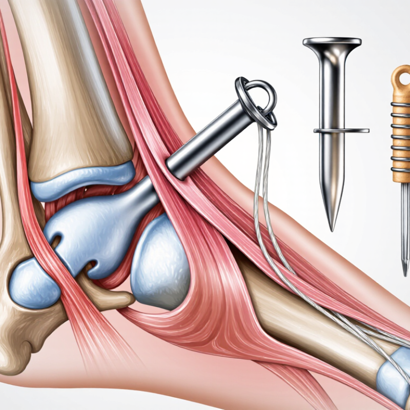
| Lateral ankle ligament repair | One or more small-diameter suture anchors placed at the ligament’s anatomical insertion. Knotless or knotted constructs may be used according to tissue quality and surgeon preference. | - Restore the native origin and insertion of the anterior talofibular and, when indicated, calcaneofibular ligament.
- Avoid the ankle joint, nearby cartilage, and the course of the superficial peroneal and sural nerves.
- Prepare the bone surface adequately without excessive removal of cortical bone.
| - Direct restoration of ligament tension and attachment.
- Small incision and limited implant prominence.
- Useful when ligament tissue is repairable.
| - Residual instability if the anchor or ligament is positioned improperly.
- Anchor pullout, suture breakage, stiffness, or irritation from prominent knots.
- Injury to cutaneous nerves or peroneal tendons is uncommon but clinically important.
| - Short-term protection in a brace or splint is commonly required.
- Progressive range of motion, weight bearing, and proprioceptive rehabilitation should follow tissue healing.
- Return to pivoting sports depends on strength, balance, and stability rather than time alone.
| High priority: anatomical accuracy, adequate bone stock, soft-tissue quality, and construct strength. |
| Medial ankle ligament repair | Suture-anchor repair or augmentation of the deltoid ligament, often using multiple fixation points when the injury involves more than one ligament layer. | - Identify the anatomical attachment on the medial malleolus and talus.
- Protect the posterior tibial tendon, saphenous nerve, and adjacent neurovascular structures.
- Check the ankle mortise and associated fractures before isolated ligament fixation.
| - Can restore medial restraint when tissue quality is sufficient.
- Allows controlled tensioning of superficial and deep ligament components.
- May be combined with fracture fixation or tendon procedures when clinically indicated.
| - Over-tensioning may restrict eversion or alter ankle mechanics.
- Medial wound problems, nerve irritation, anchor loosening, and persistent instability are possible.
- Unrecognized syndesmotic or bony injury may lead to treatment failure.
| - Use protected weight bearing when repair protection or associated injury requires it.
- Monitor swelling, wound condition, and neurovascular status.
- Rehabilitation should restore controlled ankle motion before impact activity.
| High priority: injury pattern, ligament-layer involvement, bone quality, and avoidance of excessive tension. |
| Syndesmotic stabilization | Suture-button or screw-based fixation may be selected; a suture anchor alone is generally not the standard construct for an unstable syndesmosis. | - Confirm instability with clinical examination and imaging.
- Fixation should restore fibular position within the incisura without malreduction.
- Assess associated ankle fractures and deltoid ligament injury.
| - Dynamic fixation can permit limited physiological motion during healing.
- Construct choice can be tailored to fracture pattern, instability, and bone quality.
| - Malreduction is associated with pain, altered mechanics, and poorer outcomes.
- Possible complications include hardware irritation, loss of fixation, infection, and recurrent widening.
- Incorrect drilling trajectory may damage the fibula, tibia, or nearby soft tissues.
| - Weight-bearing progression should reflect fixation stability and associated fractures.
- Follow-up imaging may be needed to assess alignment and hardware position.
- Rehabilitation should emphasize gradual restoration of ankle and lower-leg control.
| Do not substitute automatically: choose fixation based on demonstrated syndesmotic instability rather than the availability of an anchor. |
| Osteochondral lesion or cartilage-related fixation | Small, low-profile fixation may be considered only for selected fragment or soft-tissue stabilization procedures; anchor suitability depends on lesion location and bone depth. | - Use imaging and arthroscopic visualization to avoid penetrating the talar dome or opposing cartilage.
- Confirm that the anchor will be fully seated below the articular surface.
- Respect the limited bone dimensions of the talus.
| - Can provide localized fixation while limiting bulk in a joint environment.
- May support repair of selected cartilage-associated soft tissues.
| - Intra-articular prominence can cause mechanical symptoms and cartilage damage.
- Loss of fixation, synovitis, chondral injury, and persistent pain may occur.
| - Limit loading according to the repaired tissue and lesion size.
- Use a staged motion and strengthening plan to reduce shear across the repair.
- Investigate recurrent catching, locking, or swelling promptly.
| Very high priority: low profile, controlled insertion depth, and precise imaging or arthroscopic guidance. |
| Tendon or retinacular repair | Small suture anchors or transosseous sutures may be used for peroneal retinaculum, Achilles-region, or other tendon-related repairs, depending on anatomy and tissue loading. | - Place fixation in strong cortical or cancellous bone while avoiding tendon gliding surfaces.
- Restore tendon position without excessive compression or constriction.
- Consider tendon excursion and the risk of postoperative adhesions.
| - Provides direct tendon-to-bone fixation.
- Can reduce the need for larger exposure in selected procedures.
- Allows suture placement tailored to tendon thickness and tear configuration.
| - Repair elongation, rerupture, adhesions, and restricted tendon glide.
- Anchor prominence may irritate the tendon or surrounding footwear-sensitive tissue.
- Infection and wound-healing problems are possible.
| - Protect the repair from early resisted contraction and excessive stretch.
- Advance motion and strengthening according to tendon healing and clinical examination.
- Use footwear modification when swelling or local prominence causes irritation.
| High priority: tendon load, tissue thickness, anchor prominence, and rehabilitation compliance. |
| Revision repair or poor bone quality | Larger or alternative fixation may be necessary; options can include multiple anchors, bone tunnels, graft augmentation, or reconstruction rather than a single anchor. | - Evaluate previous tunnels, retained implants, bone loss, cysts, and cortical integrity.
- Avoid placing a new anchor into compromised bone or directly overlapping a prior tunnel.
- Consider advanced imaging when bone stock or alignment is uncertain.
| - Allows fixation strategy to be adapted to compromised anatomy.
- Augmentation may improve load sharing when native tissue cannot hold sutures reliably.
| - Higher risk of fixation failure, recurrent instability, infection, stiffness, and wound complications.
- Multiple implants can increase local irritation and complicate future surgery.
| - Protection is often longer and more individualized than after a primary repair.
- Document radiographic alignment and monitor for delayed healing.
- Address modifiable risks such as smoking, uncontrolled diabetes, and poor nutrition.
| Highest planning priority: bone stock, prior surgery, tissue quality, patient risk factors, and the need for reconstruction. |